Renal Imaging
Renal Imaging
Chronic Kidney Disease (CKD) is progressive condition where kidneys gradually lose function over time. Unlike acute kidney injury (AKI), CKD develops slowly and often goes unnoticed until significant damage has occurred.
Modalities of Renal Imaging
A. Ultrasound (US)
Types of Renal Ultrasound
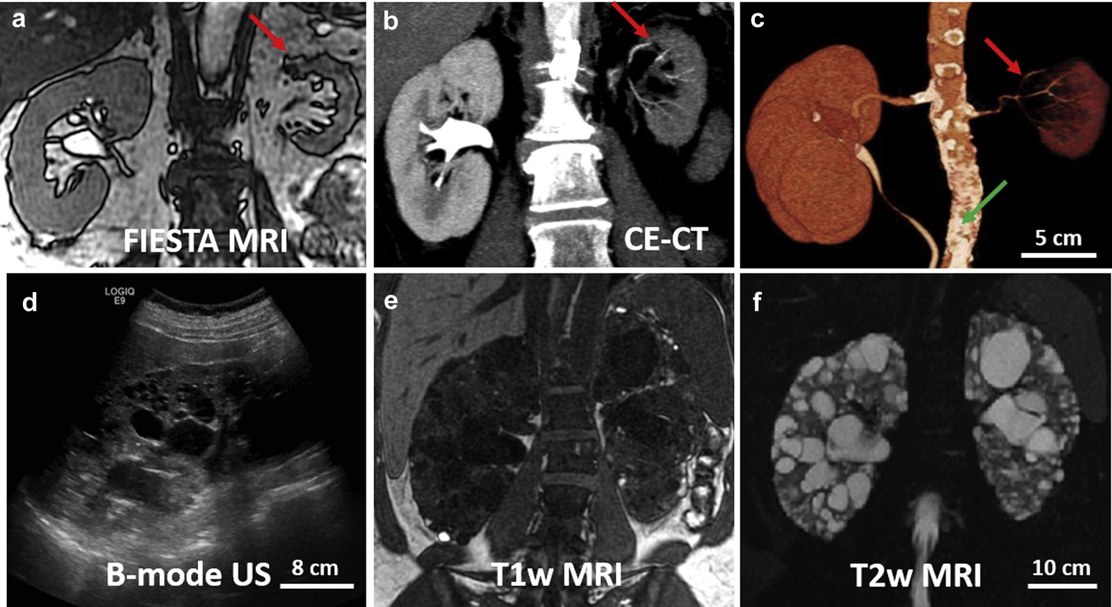
- B-mode (Brightness-mode): Standard 2D grayscale imaging for assessing kidney size, shape, and echotexture.
- Doppler Ultrasound: Evaluates blood flow in renal arteries and veins, detecting stenosis or thrombosis.
- Contrast-Enhanced Ultrasound (CEUS): Uses microbubble contrast agents to improve vascular and parenchymal visualization.
Advantages:
- Non-invasive, radiation-free, cost-effective.
- Ideal for pediatric and pregnant patients.
- Detects cysts, hydronephrosis, and renal masses.
Limitations:
- Operator-dependent.
- Limited in obese patients due to sound wave attenuation.
B. Computed Tomography (CT)
Types of Renal CT:
- Non-Contrast CT: Detects renal calculi (kidney stones), calcifications, and hemorrhage.
- Contrast-Enhanced CT (CECT):
- Corticomedullary Phase (25-40 sec): Highlights renal arteries and cortex.
- Nephrographic Phase (80-120 sec): Optimal for parenchymal lesions (tumors, infections).
- Excretory Phase (5-10 min): Visualizes collecting system and ureters.
- CT Angiography (CTA): Assesses renal artery stenosis or aneurysms.
Advantages:
- High-resolution, rapid acquisition.
- Excellent for trauma, tumors, and vascular assessment.
Limitations:
- Ionizing radiation exposure.
- Nephrotoxic contrast risk in CKD patients.
C. Magnetic Resonance Imaging (MRI)
Types of Renal MRI:
- T1/T2-Weighted Imaging: Evaluates anatomy and tissue characteristics.
- Diffusion-Weighted Imaging (DWI): Detects ischemia and tumors.
- MR Angiography (MRA): Non-invasive vascular imaging.
- Functional MRI (fMRI): Assesses renal perfusion and filtration.
Advantages:
- No ionizing radiation.
- Superior soft-tissue contrast.
- Safe for contrast-allergic patients (gadolinium alternatives available).
Limitations:
- Expensive, longer scan time.
- Contraindicated in patients with metallic implants.
D. Nuclear Medicine Imaging
Common Renal Nuclear Scans:
- DMSA Scan (Dimercaptosuccinic Acid): Assesses cortical scarring (e.g., in pyelonephritis).
- DTPA/MAG3 Scan: Evaluates glomerular filtration rate (GFR) and renal plasma flow.
- FDG-PET/CT: Detects metastatic renal cell carcinoma.
Advantages:
- Provides functional data.
- Useful in obstruction and transplant evaluation.
Limitations:
- Low spatial resolution.
- Radiation exposure.
E. Intravenous Urography (IVU) / Pyelography
Principle:
Uses iodinated contrast to visualize the urinary tract via X-rays
Applications:
Detects ureteral obstructions, strictures, and congenital anomalies.
Declining Use:
Largely replaced by CT urography due to better resolution and fewer complications.